|
|

|
Beetle morphology | ||||||||||||||||||||||||
Text © Christoph Benisch, 2007 | ||||||||||||||||||||||||
1. Basic scheme of beetle morphology | ||||||||||||||||||||||||
|
The morphology of the beetle follows a quite uniform scheme. In the diagram on the left side
the basic morphology of a beetle is shown (Carabus auratus, dorsal view). A typical characteristic of
beetles are the hard elytra. The scientific (Latin) name of the order is Coleoptera,
which translates to "sheathed wings".
The abdomen is the 3rd main segment of the body. The upper side is covered by the
elytra, the lower side forms the venter. The abdomen consists of 8-9 segments. The dorsal segments
are called tergite, the ventral segments sternite. The segments are more heavily chitinized and
flexibly connected to each other, allowing the abdomen more flexibility than the head and thorax.
The apical tergite is usually visible in dorsal view and is called pygidium. The apical sternite is
also called anal sternite. At sides of the tergites tiny holes are located, the so called spiracles
Through these pores oxygen-rich air can diffuse into tracheal tubes. | ||||||||||||||||||||||||
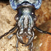
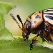
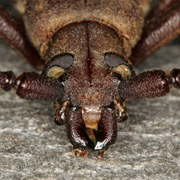
2. Head and mouthparts | ||||||||||||||||||||||||
|
The head is more or less flexibly attached to the thorax by
the cervix. The head is composed of a rigid capsule and contains the eyes (oculi), antennae and
mouthparts. On the sides of the head below/in front of the eyes the cheeks (genae) are located.
Those parts located on the side behind the eyes are called temple (tempus). The upper side of the
head in front/between the eyes is the forehead (frons), the upper part posterior to that is called
vertex.
The front part of the head is called clypeus and is often separated from
the head by a rigid suture, the frontoclypeal suture. Attached to the clypeus is the upper lip (labrum),
which is separated from the clypeus by the somewhat flexible clypeolabral suture. In some beetle species,
the labrum is covered by the clypeus and not visible in dorsal view. On the anterior end of the head the
mouthparts are located, which consist of several parts. Species feeding on plants use their mandibles to
bite off and chew their food, predators usually have edged and pointy mandibles to capture and retain their prey.
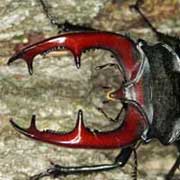
In some species, the mandibles are not functional for food intake, e.g. in the males of the stag beetle Lucanus
cervus. Their mandibles are formed like antlers, which are used for fights between male opponents.
On the lower front side of the head the maxilla are located. At their sides segmented palps (palpus maxillaris)
are attached. They are usually visible from dorsal view. The labium or lower lip finally is a fused structure
consisting of several parts (submentum, mentum, glossa). The segmented palps attached to the mentum are called
palpus labialis.
| ||||||||||||||||||||||||
3. Antennae | ||||||||||||||||||||||||
|
The antennae vary greatly among the beetles and are of high
relevance for the determination of many species. The serve for sensory perception and can detect
motion, odor and chemical substances. They are segmented and usually consist of 11 parts, the first part
is called scape (scapus), the 2nd pedicelle (pedicellus). The other segments are jointly called
flagellum. There are many different types of antennae in beetles. Antennae with a thread-like shape are called
filiform [A], gradually clubbed antenna are called clavate [B], such abruptly clubbed at the end are called
capitate [C]. Antennae with a saw-toothed shape are called serrate [D], antenna formed like a comb are
called pectinate [E]. If the first segment is longer and the antenna is bent, it is called geniculate [F].
Antennae ending in nested plates are known as lamellate [G]. | ||||||||||||||||||||||||
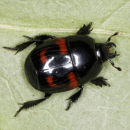
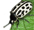
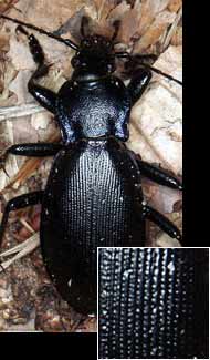
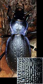
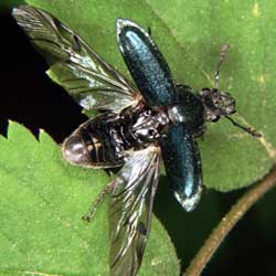
4. Elytra and alae | ||||||||||||||||||||||||
|
The elytra are the stiff and strongly sclerotized forewings of
the beetles, modified to protect the sensitive hindwings (alae) when at rest.
They are not any longer used for flying. In most cases, the elytra cover the abdomen, however
there are beetle species with shortened elytra (e.g. rove beetles (Staphylinidae)), in which
the tergites are at least partially visible. The elytra meet along the elytral suture,
a distinctive longitudinal line across the abdomen. Adjacent to the elytral suture in many cases
a sharp thin line can be observed, the so called stria suturalis. In some species the elytra are fused
together and form a closed carapace. In those cases also the membranous wings (alae) are reduced or totally
missing and these are flightless (e.g. some ground beetles).
| ||||||||||||||||||||||||
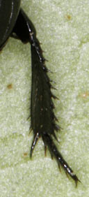
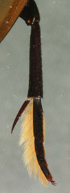
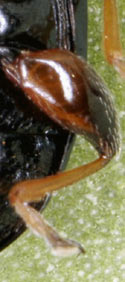
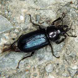
5. Legs | ||||||||||||||||||||||||
The beetles, like all other insects have 3 pairs of legs. One pair
of legs is anchored in the pro-, meta- and mesothorax. The leg is segmented in six parts:
The coxae are articulations, anchoring the leg firmly into the thorax, yet allowing front-and-back movement.
The trochantin is attached to the coxae and often barely visible. The next segment is the thigh, also called femur.
At the apical end of the femur the shin (tibia) is attached. The last segment is called foot (tarsus) and
consists of 2-5, mostly 4-5 parts. They can be broadened and carry sticky pads of packed setae (e.g. in males of
ground beetles or in longhorn beetles).
| ||||||||||||||||||||||||


 For the determination of several orders of insects, e.g.
hymenopterans and dipterans the veins are highly relevant, however in the determination of beetles
the alae and their vein structure are used very seldom. The basic principle of a membranous beetle
wing is illustrated in the diagram on the left. The costal vein (C) is the front vein and is located
on the front edge of the wing. This vein is unbranched. Behind the costal vein the subcostal vein (Sc)
is located, which ends at the edge of the wing as well. The following longitudinal veins are called
radial vein (radius, R), median vein (media, M), cubital vein (cubitus, C) and anal vein (analis, A).
These veins are branched. Together with the transversal veins they enclose segments, which are also
referred to as cell. Those cells enclosed by veins on all sides are called closed cell, those located
at the edge of the wing are called open cell.
For the determination of several orders of insects, e.g.
hymenopterans and dipterans the veins are highly relevant, however in the determination of beetles
the alae and their vein structure are used very seldom. The basic principle of a membranous beetle
wing is illustrated in the diagram on the left. The costal vein (C) is the front vein and is located
on the front edge of the wing. This vein is unbranched. Behind the costal vein the subcostal vein (Sc)
is located, which ends at the edge of the wing as well. The following longitudinal veins are called
radial vein (radius, R), median vein (media, M), cubital vein (cubitus, C) and anal vein (analis, A).
These veins are branched. Together with the transversal veins they enclose segments, which are also
referred to as cell. Those cells enclosed by veins on all sides are called closed cell, those located
at the edge of the wing are called open cell.